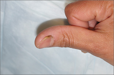
The color change in this case is subtle, but consistent with green nail syndrome also termed chloronychia.
Green nail syndrome is a form of chronic paronychia caused by Pseudomonas aeruginosa and is associated with frequent water exposure. Pseudomonas expresses pyocyanin and pyoverdine, leading to the characteristic blue or green coloration. A Woods lamp may reveal neon green fluorescence.
Risk factors include frequent water exposure, use of an instrument to relieve symptoms of an ingrown nail, and chronic immunosuppression. In this case, frequent hand washing being performed by this patient during the COVID-19 pandemic was the likely culprit.
The differential diagnosis includes simple structural onycholysis, onychomycosis, and nail psoriasis. Polymicrobial infection can occur. When in doubt, culture or pathologic examination of nail clippings with Grocott methenamine silver and periodic acid–Schiff can identify coincident fungal infection.1 While systemic pseudomonas infection could also occur with green nail syndrome, the latter is usually only a mild nuisance and not associated with severe disease.
Treatment consists of avoidance of water and instrumentation. Topical sodium hypochlorite 2% solution applied to the nail or acetic acid soaks for 2 to 3 weeks is often curative. Antibiotic therapy may also be used. Topical tobramycin or gentamicin are available as otic or ophthalmic drops and can be applied just under the nail.
The patient was advised to meticulously dry her hands after washing them and to apply 1 drop of topical gentamicin solution 0.3% tid for 3 weeks. The green discoloration cleared in 3 weeks and the onycholysis resolved within 4 months.
Text courtesy of Jonathan Karnes, MD, medical director, MDFMR Dermatology Services, Augusta, ME. Photos courtesy of Jonathan Karnes, MD (copyright retained).


